How does a dental x-ray machine work?
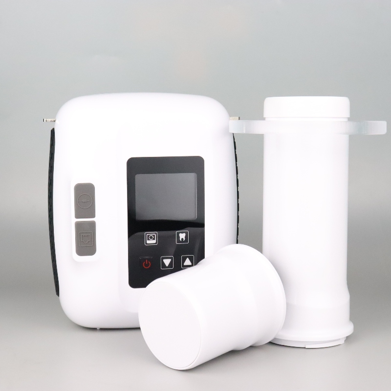
Dental X-ray machines, also known as dental X-ray units, are imaging devices used for diagnostic purposes in oral medicine. They operate based on specific principles to generate images of teeth, gums, and jaws, aiding doctors in accurately diagnosing oral diseases. Below is a detailed explanation of the operating principles of dental X-ray machines:
I. Main Components
Dental X-ray machines primarily consist of the following components:
X-ray Generator: Used to produce high voltage and high current to drive the X-ray tube to generate X-rays.
X-ray Tube: Composed of a cathode and an anode, with the cathode containing a rotating tungsten target and the anode being a metal cylinder. When high voltage and high current generated by the X-ray generator act on the X-ray tube, they stimulate the cathode to produce high-energy electrons, which collide with the tungsten target of the anode to produce X-rays.
Detector/Imaging Plate: Used to receive X-rays and convert them into visible light images or digital signals.
Image Processing System: Processes the signals received by the detector to generate clear digital images.
Display: Used to display the processed digital images for doctors to observe and analyze.
II. Operating Process
The operating process of dental X-ray machines is as follows:
X-ray Production: When the X-ray generator produces high voltage and high current, these energies act on the X-ray tube, stimulating the cathode to produce high-energy electrons. These electrons accelerate under the action of an electric field and collide with the tungsten target of the anode, releasing X-rays.
X-ray Penetration: The generated X-rays penetrate the patient's teeth, gums, jaws, and other tissues. Since different tissues absorb X-rays to varying degrees, different images can be formed on the detector.
Image Conversion: After penetrating the tissues, the X-rays are received by the detector. The detector converts the received X-rays into visible light images or digital signals. For digital dental X-ray machines, the detector is usually an imaging plate capable of converting X-rays into digital signals.
Image Processing: The digital signals received by the detector are sent to the image processing system for processing. The image processing system enhances, filters, and processes the signals to generate clear, high-resolution digital images.
Image Display: The processed digital images are sent to the display for viewing, allowing doctors to diagnose oral diseases by observing the images on the display.
III. Applications and Advantages
Dental X-ray machines have a wide range of applications in oral medicine diagnostics, such as capturing X-ray images of teeth and jaws to help doctors diagnose oral diseases like cavities, periodontal disease, wisdom teeth issues, and more. Additionally, they can be used for surgical navigation and orthodontic treatment. Dental X-ray machines offer various exposure times and focal lengths, allowing for selection based on different diagnostic needs. Furthermore, due to their use of low-dose X-rays, the impact on patients' health is minimal.
In summary, dental X-ray machines, through their unique operating principles, are capable of generating clear, high-resolution oral images, providing powerful support for the diagnosis and treatment of oral diseases.
